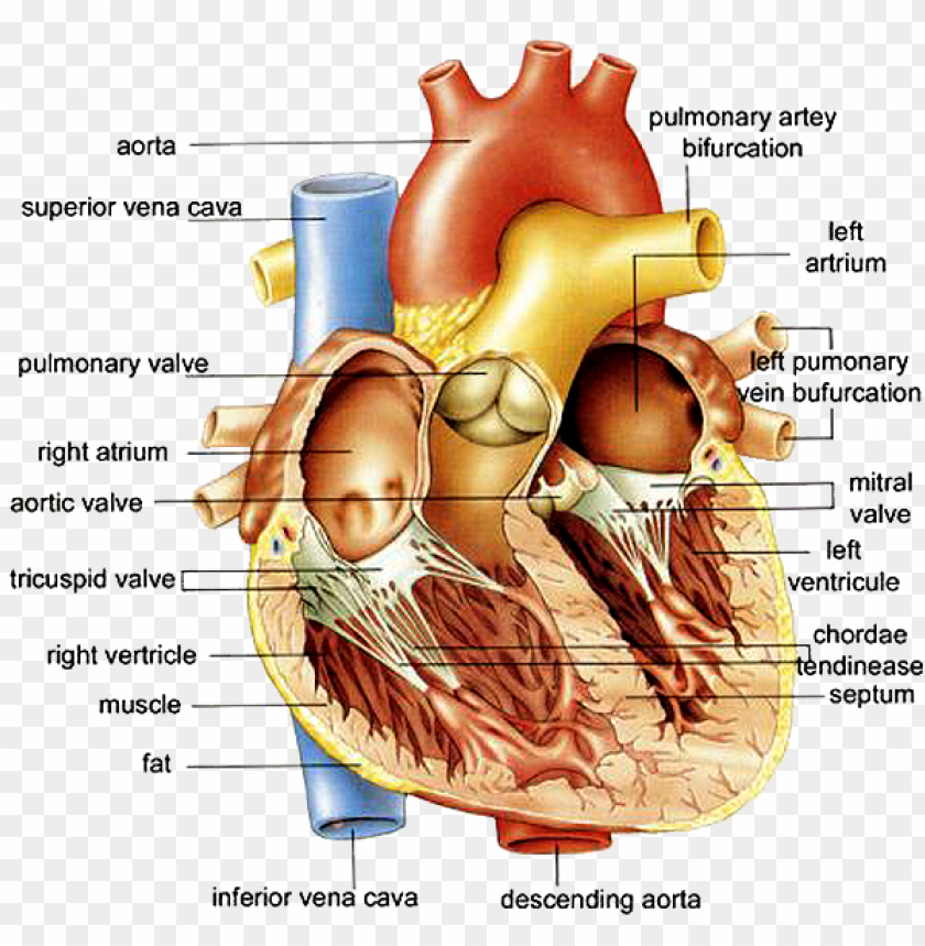
40 human heart diagram with labels
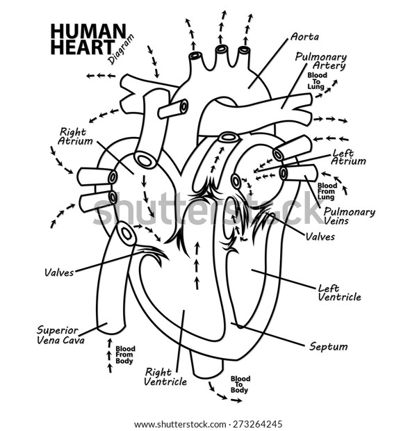
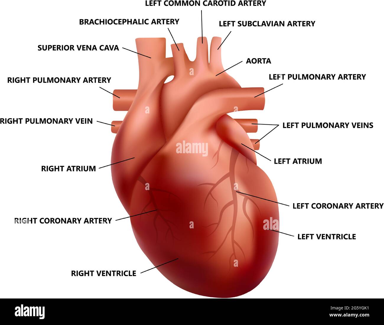
› heart › picture-of-the-heartHuman Heart (Anatomy): Diagram, Function, Chambers, Location ... Cardiomyopathy: A disease of heart muscle in which the heart is abnormally enlarged, thickened, and/or stiffened. As a result, the heart's ability to pump blood is weakened. As a result, the heart ... byjus.com › biology › human-heartHuman Heart - Anatomy, Functions and Facts about Heart - BYJUS The human heart is one of the most important organs responsible for sustaining life. It is a muscular organ with four chambers. The size of the heart is the size of about a clenched fist. The human heart functions throughout a person’s lifespan and is one of the most robust and hardest working muscles in the human body.
anatomysystem.comAnatomy System - Human Body Anatomy diagram and chart images ... Cell Picture With Labels Image Diagram - Cell Picture With Labels Image Chart - Human anatomy diagrams and charts explained. This anatomy system diagram depicts Cell Picture With Labels Image with parts and labels. Best diagram to help learn about health, human body and medicine.

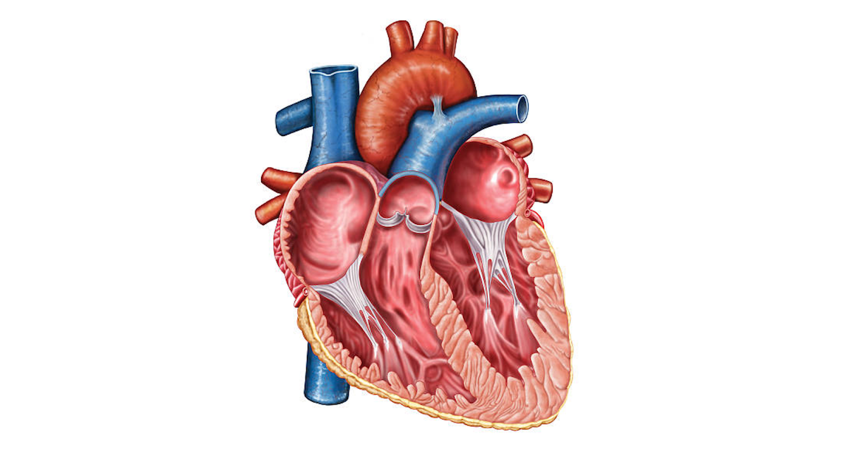
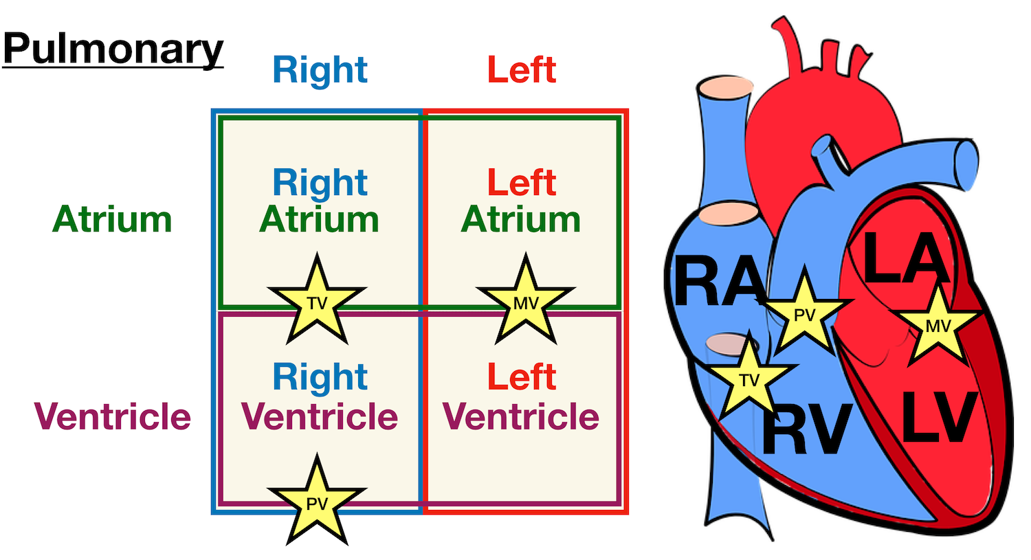
Human heart diagram with labels
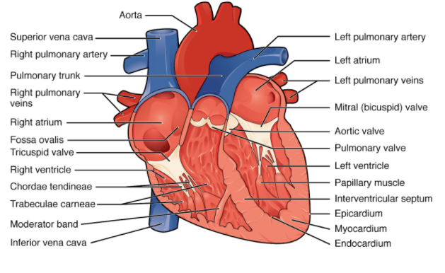
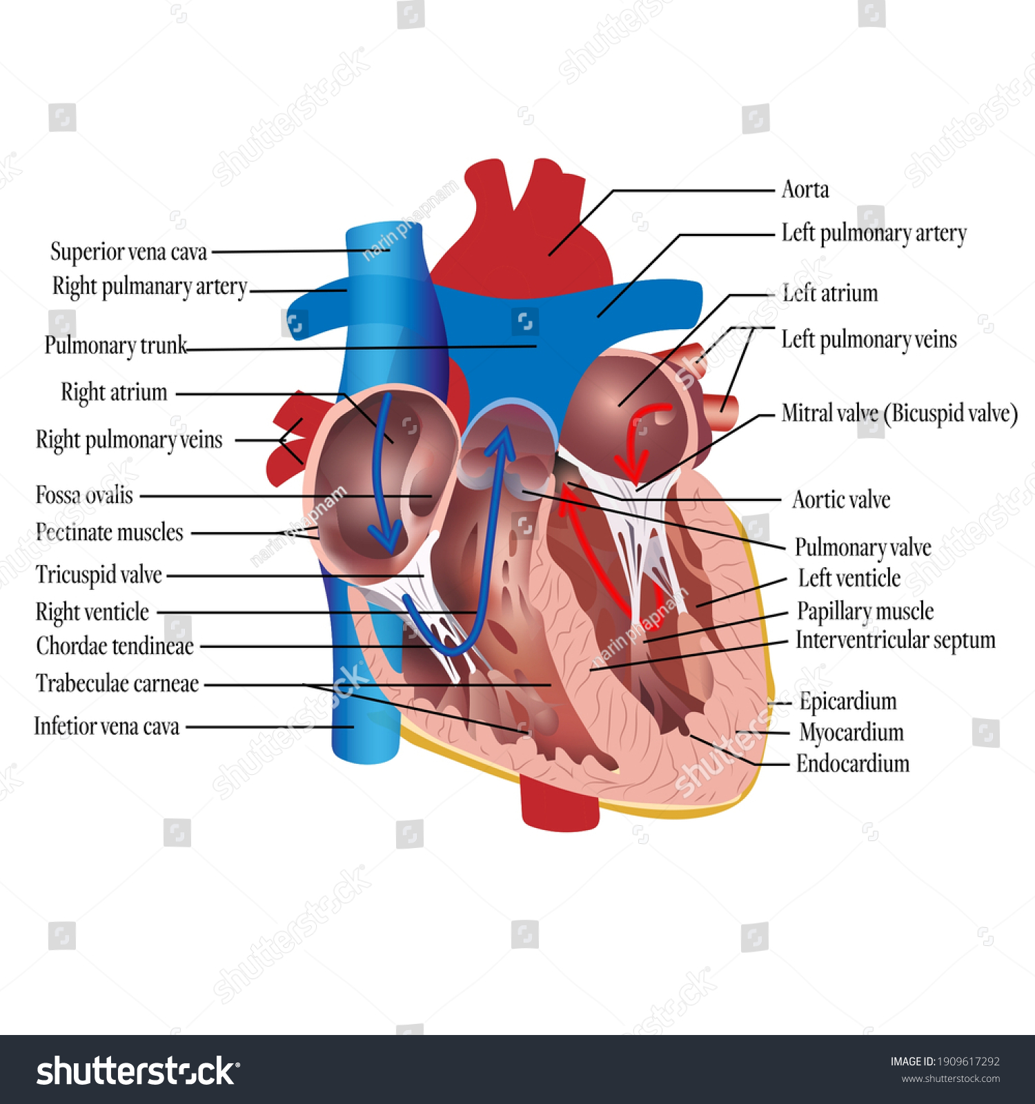
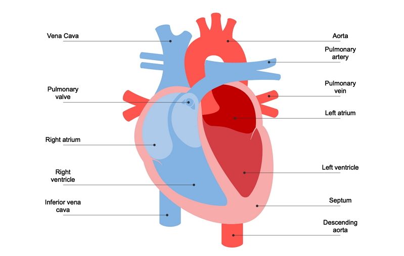
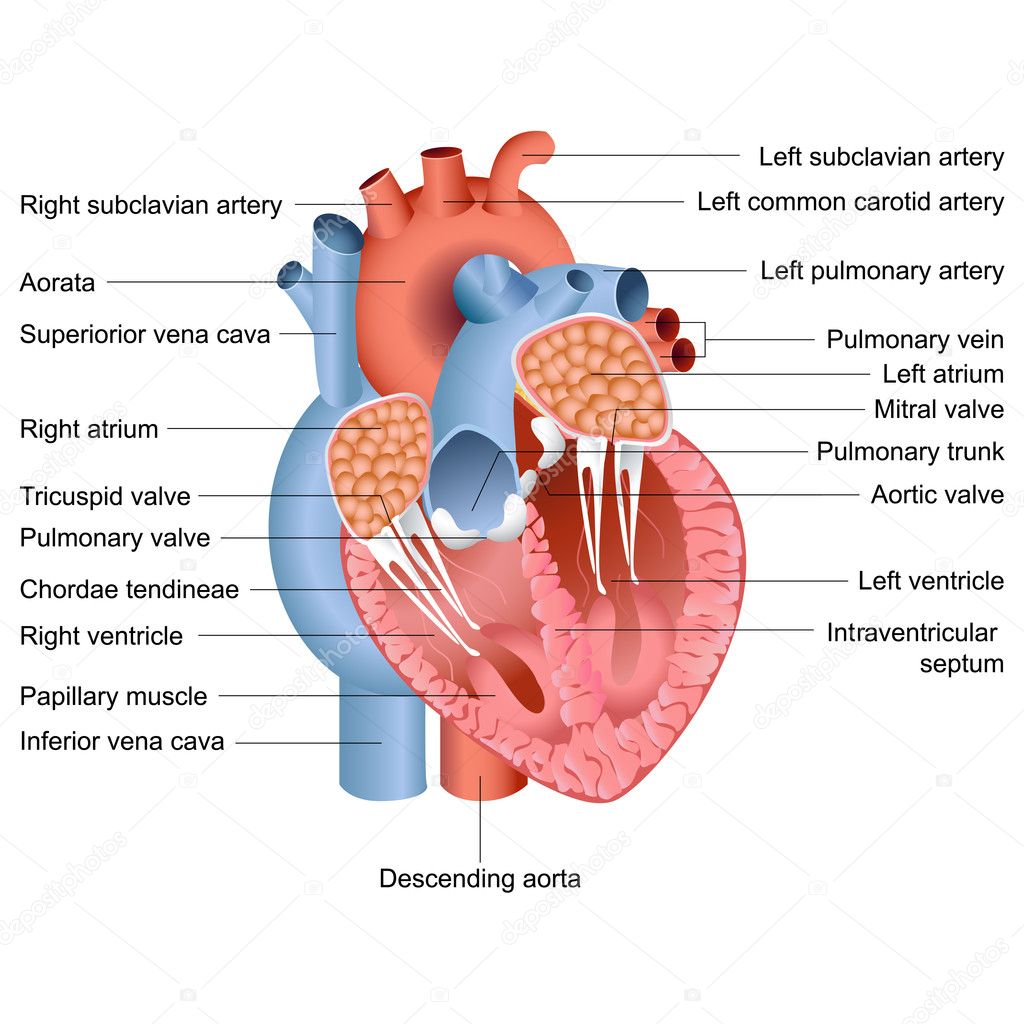
en.wikipedia.org › wiki › File:Heart_diagram-enFile:Heart diagram-en.svg - Wikipedia You are free: to share – to copy, distribute and transmit the work; to remix – to adapt the work; Under the following conditions: attribution – You must give appropriate credit, provide a link to the license, and indicate if changes were made. › Draw-a-Human-HeartHow to Draw a Human Heart: An Easy Step-By-Step Guide - wikiHow Sep 20, 2022 · The heart works like a pump and beats 100,000 times a day. The heart has two sides, separated by an inner wall called the septum. The right side of the heart pumps blood to the lungs to pick up oxygen. The left side of the heart receives the oxygen-rich blood from the lungs and pumps it to the body. commons.wikimedia.org › wiki › File:Diagram_of_theFile : Diagram of the human heart (cropped).svg - Wikimedia Sep 29, 2022 · English: Diagram of the human heart 1. Superior vena cava 2. 4. Mitral valve 5. Aortic valve 6. Left ventricle 7. Right ventricle 8. Left atrium 9. Right atrium 10. Aorta 11. Pulmonary v
Human heart diagram with labels. › science › answerHuman Body Worksheets - Easy Teacher Worksheets A human body, like a machine, is made of numerous structures that work together to perform a specific function. These structures are categorized as cells, tissues, organs, and organ systems. The systems of the human body include the digestive, nervous, and other major structures that work interdependently to ensure proper functioning. commons.wikimedia.org › wiki › File:Diagram_of_theFile : Diagram of the human heart (cropped).svg - Wikimedia Sep 29, 2022 · English: Diagram of the human heart 1. Superior vena cava 2. 4. Mitral valve 5. Aortic valve 6. Left ventricle 7. Right ventricle 8. Left atrium 9. Right atrium 10. Aorta 11. Pulmonary v › Draw-a-Human-HeartHow to Draw a Human Heart: An Easy Step-By-Step Guide - wikiHow Sep 20, 2022 · The heart works like a pump and beats 100,000 times a day. The heart has two sides, separated by an inner wall called the septum. The right side of the heart pumps blood to the lungs to pick up oxygen. The left side of the heart receives the oxygen-rich blood from the lungs and pumps it to the body. en.wikipedia.org › wiki › File:Heart_diagram-enFile:Heart diagram-en.svg - Wikipedia You are free: to share – to copy, distribute and transmit the work; to remix – to adapt the work; Under the following conditions: attribution – You must give appropriate credit, provide a link to the license, and indicate if changes were made.
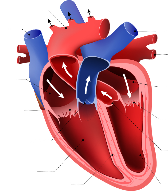
blank heart blood flow - Clip Art Library
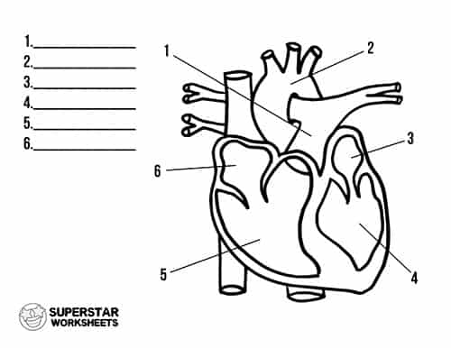
Heart Worksheets - Superstar Worksheets

The Human Heart Labeled Stock Photo, Picture And Royalty Free ...
Draw a labelled diagram of the internal structure of class 11 ...

Anatomy of the human heart stock illustration. Illustration ...

Simple heart diagram | Simple heart diagram labeled | Human ...
poster of human heart anatomy with hand written labels of the ...

Information poster human heart diagram Royalty Free Vector
http - //www - anatomybox - com/wp heart diagram - human ...
1: Labeled illustration of the human heart 1 [1]. This figure ...
Human Heart Diagram Anatomy Stock Vector (Royalty Free ...

Circulatory System Heart Labeled Stock Illustrations – 24 ...

Diagram of a human heart. | Download Scientific Diagram
Anatomy Human Heart Cross Sectional Diagram Stock Vector ...
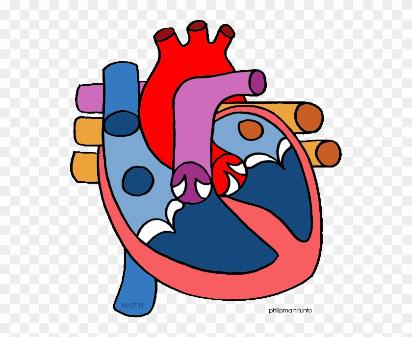
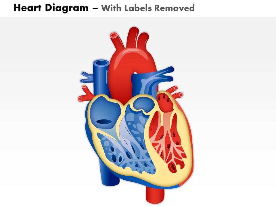
Human Heart Clipart - Heart Diagram No Labels - Free ...

The Human Heart Diagram | Quizlet

Human Heart Diagram Labeled | Science Trends

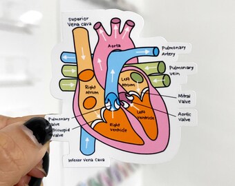
Free Printable Human Heart Diagram for Kids – Labeled and ...
Realistic heart anatomy with descriptions. Diagram of human ...
Understanding Human Heart with Heart Diagram | EdrawMax Online

Heart Anatomy Stock Illustration - Download Image Now ...
Label the Human Heart | eCampusOntario H5P Studio

Diagram of a human heart. | Download Scientific Diagram

Human Heart Diagram Anatomy Diagram Educational Chart Cool Wall Decor Art Print Poster 12x18
Free Heart Diagram Unlabeled, Download Free Heart Diagram ...
Heart Anatomy: Labeled Diagram, Structures, Blood Flow ...

How to draw internal structure of Human heart - Easy version ...

Simple heart diagram | Simple heart diagram labeled | Human ...

File:Heart diagram-en.svg - Wikimedia Commons
Heart diagram Vector Art Stock Images | Depositphotos
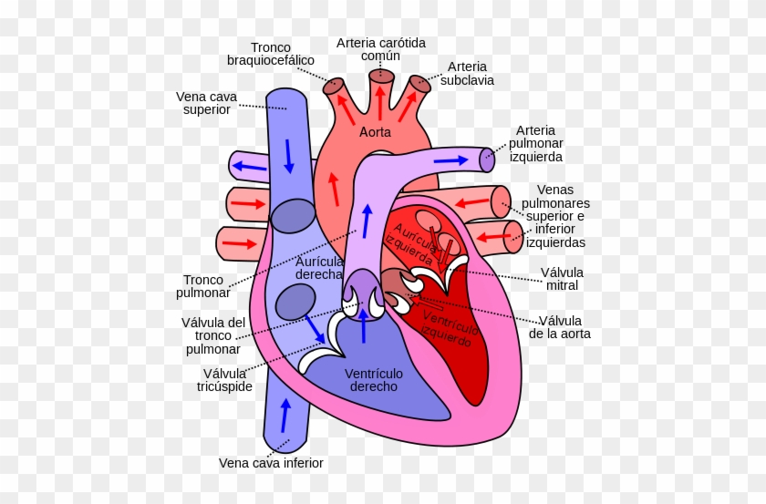
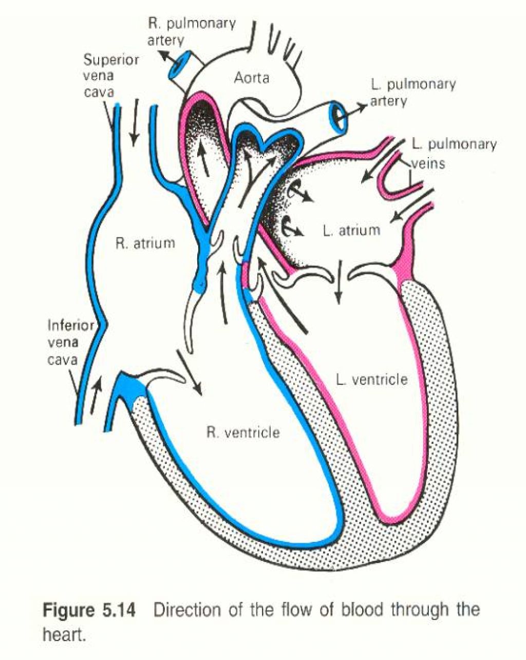
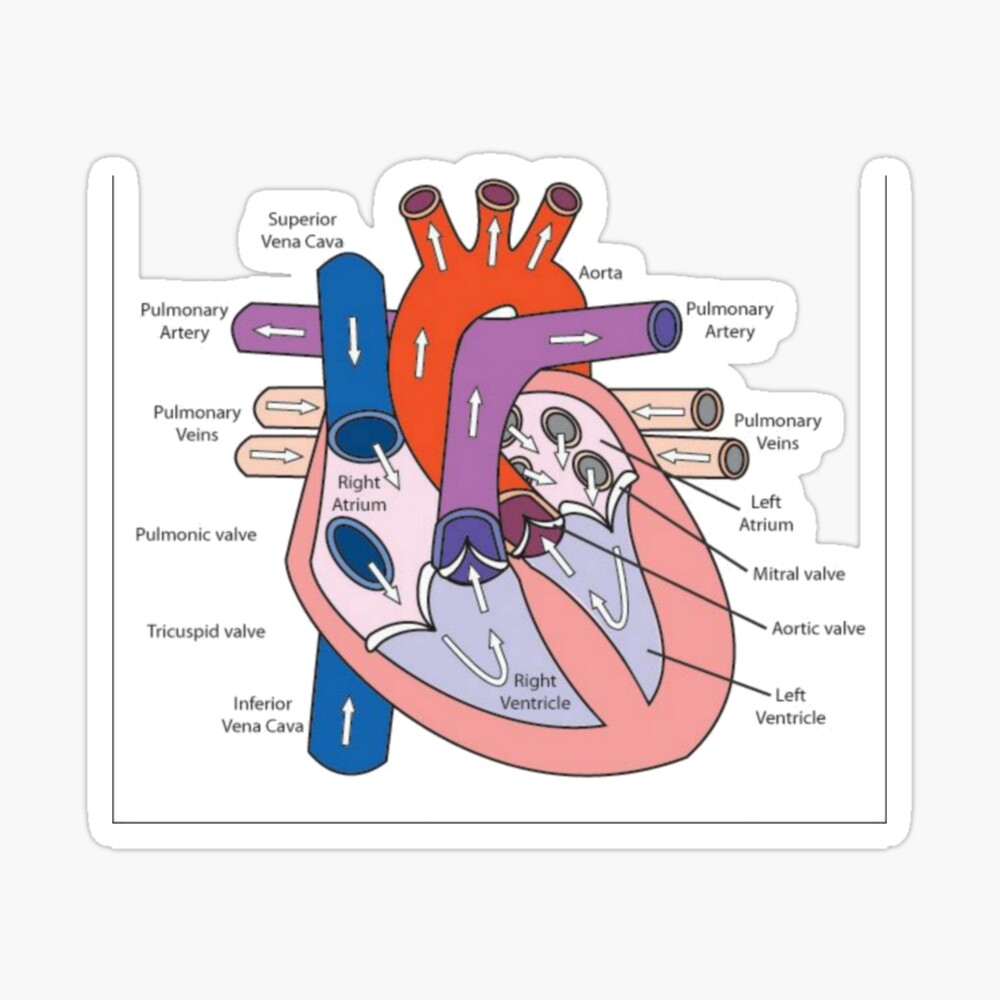
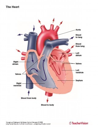
Diagram Of The Human Heart - Flow Of Blood Through The Heart ...
Heart Diagram - Etsy

File:Diagram of the human heart hu.svg - Wikipedia

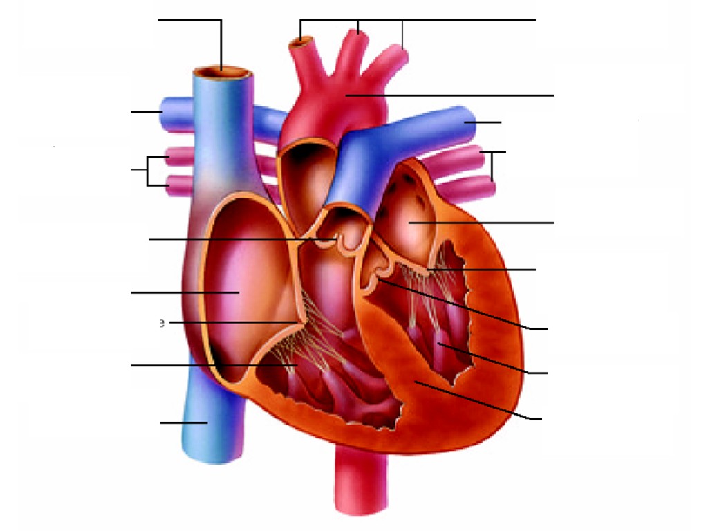
Draw the well labelled diagram of internal structure of Human ...
Heart Anatomy: Labeled Diagram, Structures, Blood Flow ...

Sketch Human Heart Vector & Photo (Free Trial) | Bigstock
0514 Heart Human Anatomy Medical Images For PowerPoint ...
Draw a labelled diagram of sectional view of human heart of ...
Anatomy of the Human Heart Printable (6th - 12th Grade ...

Draw a labelled diagram of internal structure of human heart.
Post a Comment for "40 human heart diagram with labels"