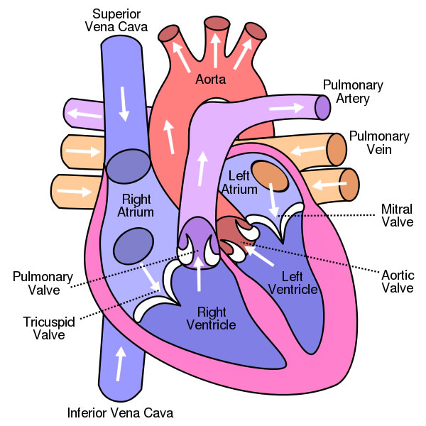
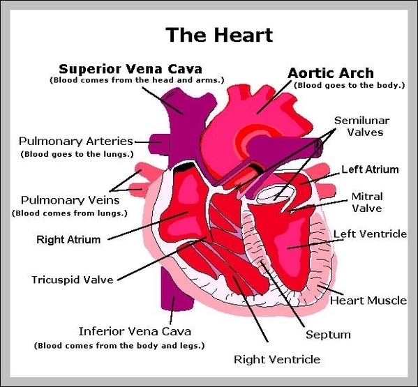
43 diagram of a human heart with labels
Eukaryotic Cell: Definition, structure and organelles | Kenhub The eukaryotic cells types are generally found in animals, plants, algae, and fungi. For the purpose of this article, the primary focus will be the structure and histology of the animal cell. The major differences between animal and plant cells will be explored as well. As previously stated, the fundamental components of a cell are its organelles. How to Count Respirations - What You Need to Know - Drugs.com This can give a false respiratory rate. Use a watch with a second hand and count his breaths for 60 seconds. Use any of the following methods to count: Look at his chest rise and fall. One rise and one fall are counted as 1 breath. Listen to his breaths. Place your hand on the person's chest to feel the rise and fall.
› heart › picture-of-the-heartHuman Heart (Anatomy): Diagram, Function, Chambers, Location ... Cardiomyopathy: A disease of heart muscle in which the heart is abnormally enlarged, thickened, and/or stiffened. As a result, the heart's ability to pump blood is weakened. As a result, the heart ...

Diagram of a human heart with labels
en.wikipedia.org › wiki › File:Diagram_of_the_humanFile:Diagram of the human heart (cropped).svg - Wikipedia Diagram of the human heart, created by Wapcaplet in Sodipodi. ... Add Inferior vena cava and pericardium labels: 18:08, 14 August 2018: 656 × 631 (209 KB) Jmarchn: › male-human-anatomy-diagramMale Human Anatomy Diagram Pictures, Images and Stock Photos Pacemaker Diagram Cross section of a human heart with pacemaker fitted, showing the major arteries and veins. This is an EPS 10 vector illustration and includes a high resolution JPEG. male human anatomy diagram stock illustrations Functions of the Cardiovascular System | Healthcare-Online The heart pumps about 10 pints of blood through the body with very beat. That blood travels through a complex system of arteries, veins and capillaries. The blood carries oxygen and nutrients to every cell of the body. The cardiovascular system is actually comprised of two systems: The pulmonary circulation, in which the heart pumps the blood ...
Diagram of a human heart with labels. › print › heart-diagramsHeart Diagram – 15+ Free Printable Word, Excel, EPS, PSD ... Teachers and students use the heart diagram, in biological science, to study the structure and functions of a human being’s heart. Friends and colleagues on the other hand may find this diagram template useful when it comes to sending special, personalized gifts to their family members and significant others. Download the template today, and ... Trunk Region (Torso) < Regional Anatomy << Human Anatomy <<< Body ... Trunk (Torso) In our body, the Trunk Region (Torso), an anatomical term for the central part of the body, is a combination of both the thoracic region (chest), including the mammary region (breasts), and the abdomen region (belly region) including the naval (umbilicus region), coxal region, and pubic region. In our body, the Trunk Region (Torso) as an anatomical term for the central part of ... How Drugs Affect the Brain & Central Nervous System Drugs that can impact GABA levels: benzodiazepines. Norepinephrine: Similar to adrenaline, norepinephrine is often called the "stress hormone," as it speeds up the central nervous system in response to the "fight-or-flight" response. It also homes focus and attention while increasing energy levels. Double Helix - Genome.gov Definition. …. Double helix, as related to genomics, is a term used to describe the physical structure of DNA. A DNA molecule is made up of two linked strands that wind around each other to resemble a twisted ladder in a helix-like shape. Each strand has a backbone made of alternating sugar (deoxyribose) and phosphate groups.
commons.wikimedia.org › wiki › File:Diagram_of_theFile:Diagram of the human heart (cropped).svg - Wikimedia Aug 08, 2022 · English: Diagram of the human heart 1. Superior vena cava 2. 4. Mitral valve 5. Aortic valve 6. Left ventricle 7. Right ventricle 8. Left atrium 9. Right atrium 10. Aorta 11. Pulmonary v Library Guides: Health Statistics & Data: Datasets/Raw Data Nightingale Open Science is a platform that connects researchers with world-class medical data from health systems around the world. Datasets of electrocardiogram waveforms, x-rays and CT scans, tissue biopsy images, and more are linked to ground-truth labels. Access requires registration. Capillaries: Anatomy, Function, and Significance - Verywell Health Capillary Function. The capillaries are responsible for facilitating the transport and exchange of gases, fluids, and nutrients in the body. While the arteries and arterioles act to transport these products to the capillaries, it is at the level of capillaries where the exchange takes place. The capillaries also function to receive carbon ... Streak Plate Method: Principle, Procedure, Uses - Microbe Online T Streak. Procedure. Repeat steps 1 to 6 as per quadrant streaking. A T shape is drawn on the bottom surface of the plate using a marker. Remove the lid of the labeled agar plate just enough to insert the loop and lightly drag the loop with suspension in a zig-zag pattern in the top half of the T. (remember to stay within the region) Close the lid and flame the inoculating loop once again.
Pedigree - Genome.gov A pedigree, as related to genetics, is a chart that diagrams the inheritance of a trait or health condition through generations of a family. The pedigree particularly shows the relationships among family members and, when the information is available, indicates which individuals have a trait (s) of interest. Narration 00:00 … 13 Free Mood Tracker Printables to Understand Yourself Better In this post, we share with you 13 beautifully designed mood tracker printables to better understand your moods and feelings. Let's get to it! What You Will Learn 13 Free Mood Tracker Printables 1. Minimalist Floral Mood Tracker 2. Mandala Mood & Gratitude Tracker 3. Weekly Printable Mood Tracker Worksheet 4. Four-Month Mood Tracker 5. Parts of Human Eye and Their Functions | MD-Health.com The iris is the area of the eye that contains the pigment which gives the eye its color. This area surrounds the pupil, and uses the dilator pupillae muscles to widen or close the pupil. This allows the eye to take in more or less light depending on how bright it is around you. If it is too bright, the iris will shrink the pupil so that they ... neuron | Definition & Functions | Britannica neuron, also called nerve cell, basic cell of the nervous system in vertebrates and most invertebrates from the level of the cnidarians (e.g., corals, jellyfish) upward. A typical neuron has a cell body containing a nucleus and two or more long fibres. Impulses are carried along one or more of these fibres, called dendrites, to the cell body; in higher nervous systems, only one fibre, the axon ...
Kids' Health - Topics - Your heart | Heart for kids, Heart diagram, Heart lesson
Annelid - Wikipedia Classification and diversity. There are over 22,000 living annelid species, ranging in size from microscopic to the Australian giant Gippsland earthworm and Amynthas mekongianus (Cognetti, 1922), which can both grow up to 3 meters (9.8 ft) long to the largest annelid, Microchaetus rappi which can grow up to 6.7 m (22 ft). Although research since 1997 has radically changed scientists' views ...

Human Heart Labelled Stock Photo - Download Image Now - iStock
Vagina Pics with its anatomy - WOMS Let's begin the anatomy of the vagina with its shape and size. The anterior wall of the vagina is about eight centimeters long. And the posterior wall of the vagina is about ten centimeters long. Relations of the anterior wall The upper half of the anterior wall is related to the base of the urinary bladder. The lower half of the urethra

Anatomy/Cardiovascular System - Wiki - Scioly.org
byjus.com › biology › human-heartHuman Heart - Anatomy, Functions and Facts about Heart - BYJUS The human heart is one of the most important organs responsible for sustaining life. It is a muscular organ with four chambers. The size of the heart is the size of about a clenched fist. The human heart functions throughout a person’s lifespan and is one of the most robust and hardest working muscles in the human body.
Human Heart Diagram - Human Body Pictures - Science for Kids
byjus.com › biology › diagram-of-heartHeart Diagram with Labels and Detailed Explanation - BYJUS The human heart is the most crucial organ of the human body. It pumps blood from the heart to different parts of the body and back to the heart. The most common heart attack symptoms or warning signs are chest pain, breathlessness, nausea, sweating etc. The diagram of heart is beneficial for Class 10 and 12 and is frequently asked in the ...
13+ Heart Diagram Templates – Sample, Example, Format Download | Free & Premium Templates
Diagram of Human Heart and Blood Circulation in It Exterior of the Human Heart A heart diagram labeled will provide plenty of information about the structure of your heart, including the wall of your heart. The wall of the heart has three different layers, such as the Myocardium, the Epicardium, and the Endocardium. Here's more about these three layers. Epicardium

human heart diagram with labels
Anatomy Of The Heart - Otosection Anatomy. the heart has a somewhat conical form and is enclosed by the pericardium. it is positioned posteriorly to the body of the sternum with one third situated on the right and two thirds on the left of the midline. the heart measures 12 x 8.5 x 6 cm and weighs ~310 g (males) and ~255 g (females) relations.

Heart Diagram Quiz
Diaphragm: Function, Anatomy, and Abnormalities - Verywell Health Here's a Quick Overview of the Azygos Vein Location Your diaphragm body part spans from the front to the back. It is the floor of the thoracic cavity and the ceiling of the abdominal cavity. 2 Many body organs are near the diaphragm. Your heart, lungs, and the upper part of your esophagus (food pipe) are in the thoracic cavity above the diaphragm.

HEALTH CARE ( The topic you are looking for...): January 2010
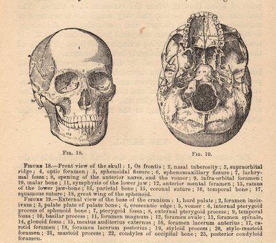
Antenatal Care Module: 6. Anatomy of the Female Pelvis and Fetal Skull The dimensions in centimetres (cm) of the pelvic inlet are shown in Figure 6.3 in both directions (top to bottom; and transverse or side to side). When you look at Figure 6.3, imagine that you are a baby in the head-down position, looking down on the pelvis from above, at the space you must squeeze through!

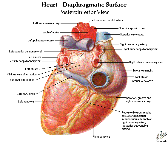
Anatomy of the Heart (Latin)
HOW TO MAKE A LUNG MODEL WITH KIDS - hello, Wonderful - Double stick tape or glue Printable template (download at my Teachers Pay Teachers Store) Instructions: Step 1. Print and cut your template out. Step 2. Trim the straws at the top. Step 3. Add some tape to the bottom to keep the two straws together. Step 4. Glue or double stick tape your nose and lips to the straws. Step 5.

Label the Heart Quiz
Heart - Wikipedia The human heart is situated in the mediastinum, at the level of thoracic vertebrae T5-T8.A double-membraned sac called the pericardium surrounds the heart and attaches to the mediastinum. The back surface of the heart lies near the vertebral column, and the front surface known as the sternocostal surface sits behind the sternum and rib cartilages. The upper part of the heart is the attachment ...

Inside Thoughts: February 2012
Positions and Functions of the Four Brain Lobes | MD-Health.com The brain is divided into four sections, known as lobes (as shown in the image). The frontal lobe, occipital lobe, parietal lobe, and temporal lobe have different locations and functions that support the responses and actions of the human body. Let's start by identifying where each lobe is positioned in the brain. Position of the Lobes

Heart Diagram Unlabeled | Medical anatomy, Medical school studying, Medical education
Hippocampus: Anatomy, functions and connections | Kenhub The hippocampus is a paired structure present in each temporal lobe of the brain. It takes its name from the Greek word for the seahorse, because it resembles this small upright-swimming fish. The hippocampus is part of a larger structure of the temporal lobe called the hippocampal formation.
heart parts – Graph Diagram
What is Amperage? (with pictures) - All the Science A current of 0.1-0.2 amps flowing through a human body is usually lethal, due to its effects on the heart. Surprisingly, with prompt treatment, victims exposed to more than 0.2 amps may survive, as the severe muscle contractions induced can protect the heart from electrical interference.
Vintage Graphic - Anatomy - Skull Diagram - The Graphics Fairy
Left-Sided and Right-Sided Heart Failure - What You Need to Know Heart failure is a condition that does not allow your heart to fill or pump properly. Heart failure may begin on the left or right side of the heart. If one side does not work properly, the other side has to work harder to function. Over time, heart failure affects both sides of the heart. Heart failure is a long-term condition that tends to ...

Heart Diagram Worksheet Blank Human Heart Worksheets Label Heart Diagram Worksheet Awesome in ...
human body | Organs, Systems, Structure, Diagram, & Facts human body, the physical substance of the human organism, composed of living cells and extracellular materials and organized into tissues, organs, and systems. Human anatomy and physiology are treated in many different articles. For detailed discussions of specific tissues, organs, and systems, see human blood; cardiovascular system; digestive system, human; endocrine system, human; renal ...
Free Unlabelled Diagram Of The Heart, Download Free Unlabelled Diagram Of The Heart png images ...
Circulatory System Diagram | New Health Advisor Heart consists of four chambers namely right atria, left atria, right ventricle and left ventricle. Both the atrium and ventricles are separated from each other with a muscular septum. Valves are present in between atria and ventricle which helps in draining the blood from upper part to lower part of the body.

The Heart Diagrams Labeled and Unlabeled
Mind Map Templates | Free to Download and Edit - Edrawsoft Step 4: Save Your Mind Map Template for Word/Excel/PowerPoint/PDF. When you finish creating your mind map in EdrawMind, one-click on the [Export] button will transfer your drawing into the editable Word/Excel/PowerPoint files or convert your mind map template into the PDF format. Thus, even non-EdrawMind users can read and edit your mind map ...

Labelled Diagram of the Human Heart - Cancer / Asbestos Cancer | Diagram of Heart | Mesothelioma >>
Human Circulatory System: Definition, Organs and Functions - Embibe Exams The human heart consists of four chambers - two ventricles and two auricles. Organs or Parts of Circulatory System The main parts of the human circulatory system include organs like blood vessels, the lymphatic system and the heart, and a fluid connective tissue called the blood. Let us now learn about the organs of the circulatory system below:
Post a Comment for "43 diagram of a human heart with labels"