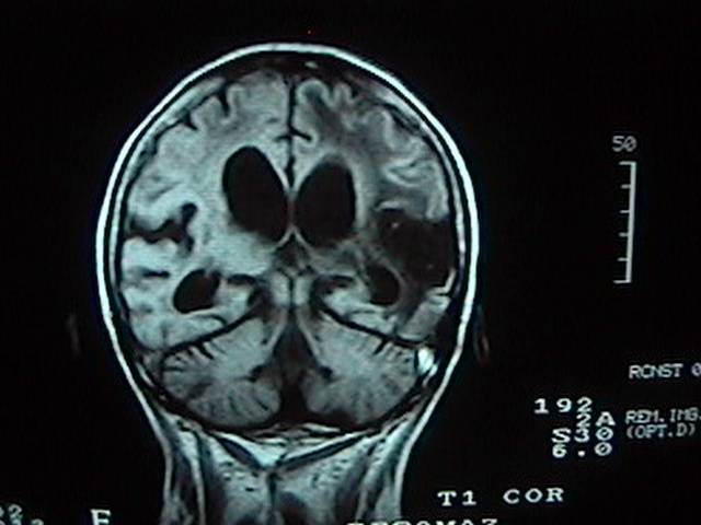
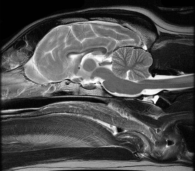
41 brain mri with labels
Vessel Wall Imaging of the Head and Neck: Approach and ... - Medscape Target Audience and Goal Statement. This activity is intended for radiologists, neuroradiologists, neurologists, and cardiologists. The goal of this activity is to educate radiologists, neuroradiologists, neurologists, and radiologic technologists about current practices in cervical and extracranial vessel wall magnetic resonance imaging (VW-MRI). What does federal law and 'the science' say about brain manipulation ... Simply labeling others as crazy or "mentally ill" for suggesting things that the person does not want discussed or even thought to be a possibility often succeeds in getting some to keep silent. It...
Primary central nervous system lymphoma | Blood - American Society of ... Primary central nervous system lymphoma (PCNSL) is an extranodal non-Hodgkin lymphoma affecting the brain, spine, cerebrospinal fluid (CSF), or vitreoretinal space. 1,2 It is a rare cancer with an annual incidence of 0.4 per 100 000 in all comers, 3,4 but the incidence increases with age to as high as 4 per 100 000 in those older than 70 years. 3 PCNSL accounts for 4% to 6% of all extranodal ...

Brain mri with labels
› 2013 › 07CPT Code for MRI Brain, Breast, Lumbar Spine and Shoulder Find below the latest Radiology CPT codes for for MRI of Brain, Breast, Lumbar Spine and Shoulder: CPT Codes for MRI Lumbar spine In human Lumbar spine is represented by the 5 vertebrae in between the ribcage and the pelvis forming the largest segment of the vertebral column. Depending on the condition that one is treated on these parts of the ... An Extension Protocol for Patients Who Previously Completed the TMS ... Protocol #8116 investigates the clinical efficacy of open-label individualized MRI-guided TMS applied to the left temperoparietal junction (TPJ) in schizophrenia patients. Participating patients who have completed the 4-week project #8116 can be screened for eligibility for this extension study in which they will continue treatment/assessment. Labelling Images - 15 Best Annotation Tools in 2022 - Folio3AI Blog RectLabel. This is another tool for image labelling, though, like LabelIMG was more comfortable with Windows, RectLabel is made for the macOS and is easy to use for all macOS users. It is free of cost and provides great ways to label images using bounding boxes and polygons.
Brain mri with labels. All You Need to Know about Chronic Microvascular Ischemic Disease Microvascular ischemic disease is a sign that there is reduced blood flow to areas of the brain. Many people who have an MRI of the brain turn out to have this condition as a result of the aging process. Arteries with age can become more rigid and narrow and less able to send blood to some of the areas. MRI sequences (overview) | Radiology Reference Article - Radiopaedia This leads to a broad categorization as follows: T1 weighted (T1W) gadolinium enhanced fat suppressed T2 weighted (T2W) fat suppressed fluid attenuated susceptibility sensitive proton density (PD) fat suppressed diffusion weighted flow sensitive MR angiography (MRA) MR venography (MRV) CSF flow studies miscellaneous › AANLIB › casesHarvard University Show labels Show list All modalities to: MR-T1 MR-T2 FDG T1/FDG T2/FDG Divergent patterns of cognitive deficits and structural brain ... Participants were labeled as either SSR or MSR based on their/their co-participant's reported sex. We identified 1,073 participants (1,037 MSR−555 cognitively unimpaired [CU]; 36 SSR−23 CU) with structural MRI data, Mini-Mental State Exam (MMSE), and Neuropsychiatric Inventory Questionnaire (NPI-Q) scores.
The Psychopathic Brain: Is It Different from a Normal Brain? There are three main parts of the brain which psychopath brain scans show significant differences in. They are in the regions of the amygdala, the prefrontal cortex, and the extended paralimbic structures. Light is shed on these regions in the paper, The Psychopath Magnetized: Insights from Brain Imaging, by Anderson and Kiehl. › en › e-AnatomyShoulder: MRI, radiographical, and illustrated anatomical ... Sep 13, 2021 · MRI of the shoulder : muscles of the rotator cuff labeled on a sagittal MR slice. An MRI of the shoulder of a healthy subject was performed in the 3 planes of space (coronal, axial, sagittal) commonly used in osteoarticular imaging, with two weightings to explore the musculoskeletal pathology of the shoulder: spin-echo T1 and proton-density ... › articles › s41586/022/04554-yBrain charts for the human lifespan | Nature Apr 06, 2022 · To extend the scope of brain charts beyond the four cerebrum tissue volumes, we generalized the same GAMLSS modelling approach to estimate normative trajectories for additional MRI phenotypes ... NEURO: Highly time-resolved 4D MR angiography using golden-angle radial ... Highly time-resolved 4D MR angiography using golden-angle radial sparse parallel (GRASP) MRI ... Share to Pinterest. Labels: Neurology : nature.com subject feeds, NeuroNature. No comments: Post a Comment. Newer Post Older Post Home. Subscribe to: Post Comments (Atom) How Changes in Length of Day Change the Brain and Subsequent Behavior. Neurons ...
SticiGui Sample Final Exam 1 - University of California, Berkeley A box contains tickets labeled with the numbers {-3, -1, 0, 1, 3}. In 100 random draws with replacement from the box, the ... to a pre-determined position in the brain (guided by MRI). Radio-frequency electricity is passed through the electrodes, to raise the temperature in the part of the brain between the electrodes to 85°C (185°F) for 90 ... Itk Snap Projects - Matlab Projects We implement ITK-SNAP a software tool for segment anatomical structures, viewing and label 3D medical images. We ensure automatic active contour segmentation pipeline with manual segmentation toolbox support. We determine ITK-SNAP matlab with full featured UI aimed at clinical researchers. Find brain mri findings | VyShows.com 14-09-2020 · MRI can detect a variety of conditions of the brain such as cysts, tumors, bleeding, swelling, developmental and structural abnormalities, infections, inflammatory conditions, or problems with the blood vessels. It can determine if a shunt is working and detect damage to the brain caused by an injury or a stroke. Ain't Nothin' Gonna Break My Strider - Razzball Fantasy Baseball Ugh..brain fart, I read your comments above on him, but that was at the beginning, so when I got to the end, I forgot. HAHAHA. Sorry! I was also excited because I had Strider yesterday..forgive me. Still, would you drop Grissom, Lindor, Adames, or Torres for him, or nah? (seems like a nah)…Thanks!
Diagnostic Imaging - Brain MRI
Twitter Finally Announces The Edit Tweet Feature; Heres The Downside ... Twitter announced the new Edit Tweet feature via a blog post. As the name suggests, the new feature allows users to edit their tweets even after it's posted. Small errors like spelling mistakes ...

Radiodiagnosis - Imaging is Amazing-Interesting cases: Phenytoin associated Cerebellar atrophy - MRI
en.wikipedia.org › wiki › Diffusion_MRIDiffusion MRI - Wikipedia Diffusion-weighted magnetic resonance imaging (DWI or DW-MRI) is the use of specific MRI sequences as well as software that generates images from the resulting data that uses the diffusion of water molecules to generate contrast in MR images.
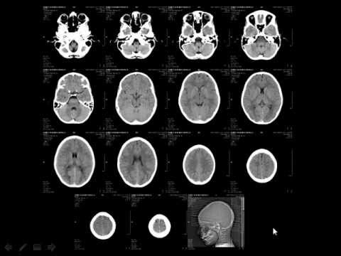
Some sample MRI images
Doctors Told Her Her Early-Onset Alzheimer's Was 'Just Stress' An initial PET scan came up inconclusive, prompting an initial diagnosis of brain fog due to stress or menopause symptoms — and the search for a second opinion. Michele credits the Alzheimer's Association with making the journey less isolating. Michele is on Aduhelm, the controversial drug that is challenging to be prescribed.
Radiology - Normal brain anatomy - CT and MRI - YouTube
Amitriptyline/antihistaminics/corticosteroids | SpringerLink The brain MRI localized the stroke in the left lateral medulla and confirmed Wallenberg syndrome. Concomitantly with the other symptoms, she developed a severe itch restricted to the right side of the head, extending to the right upper limb and trunk in the following weeks. ... off-label dupilumab was started. After 16 weeks, a reduction in PN ...

356 best MRI images on Pinterest | Radiology, Rad tech and Anatomy
Injury update on Mohammad Rizwan 00:00 / 29:33. It is understood that Rizwan underwent an MRI scan after the game, but a follow-up scan has been recommended as a precautionary measure, according to various reports. Rizwan is the leading run-scorer in Asia Cup 2022 with 192 runs, including two fifties in three matches. The opener also has 1521 T20I runs since the start of 2021 ...
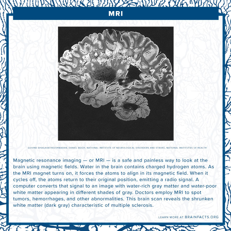
MRI
braintumorsegmentation.orgMICCAI BRATS - The Multimodal Brain Tumor Segmentation Challenge RSNA-ASNR-MICCAI Brain Tumor Segmentation (BraTS) Challenge 2021 . The Brain Tumor Segmentation (BraTS) challenge celebrates its 10th anniversary, and this year is jointly organized by the Radiological Society of North America (RSNA), the American Society of Neuroradiology (ASNR), and the Medical Image Computing and Computer Assisted Interventions (MICCAI) society.

Brain Radiology | 118.164
Internal carotid artery | Radiology Reference Article - Radiopaedia Gross anatomy Origin. The common carotid artery bifurcates to form the internal carotid and the external carotid artery (ECA).Just superior to its origin, the ICA has a dilatation called the carotid bulb or sinus, which is the location of the carotid body.. In most cases, the carotid bifurcation occurs between the levels of the C3 and C5 vertebrae, or between the levels of the hyoid bone and ...
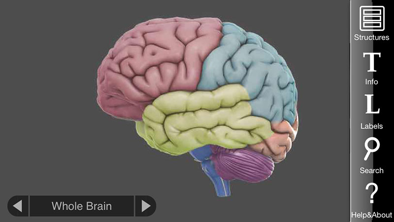
3D Brain App, an interactive way to learn about the different parts of the human brain | Apps ...
› en › e-AnatomyNormal chest MDCT with anatomic labels | e-Anatomy - IMAIOS Mar 10, 2022 · IMAIOS and selected third parties, use cookies or similar technologies, in particular for audience measurement. Cookies allow us to analyze and store information such as the characteristics of your device as well as certain personal data (e.g., IP addresses, navigation, usage or geolocation data, unique identifiers).

Neuroradiology Cases: Multiple System Atrophy MRI
Biotech Stock Roundup: BMY's Study Update, INCY's Drug Label Expansion ... The primary objective of this study was to detect a dose response for the composite endpoint of symptomatic ischemic stroke + MRI detected covert brain infarction across a 16-fold dose range ...
Sagittal Plane MRI Head Atlass
mne.viz.plot_topomap — MNE 1.2.dev0 documentation outlines 'head' | 'skirt' | dict | None. The outlines to be drawn. If 'head', the default head scheme will be drawn. If 'skirt' the head scheme will be drawn, but sensors are allowed to be plotted outside of the head circle. If dict, each key refers to a tuple of x and y positions, the values in 'mask_pos' will serve as ...

Dr Balaji Anvekar FRCR: Hypothalamic Hamartoma presenting with precocious puberty
Persistent White Matter Changes in Recovered COVID-19 Patients - Medscape Diffusion MRI was acquired with the following parameters: repetition time/echo time = 5400/92 ms, field of view = 224 × 224 mm, 112 × 112 matrix, 40 slices, 2 × 2 × 3 mm 3 voxels, bandwidth = 1654...

Neuroanatomy - encyclopedia article - Citizendium
Medical Image Processing using Matlab - MATLAB PROJECTS Overall, it is an efficient mathematical tool to get the desired results from MRI, PET, Fluorescein Angiogram, and CT images. In specific, we have given the reason behind the use of Matlab in medical image processing. Why to use Matlab for Medical Image Processing? Easy to automatically create the label the image data in the folder tags

ventricular system overview :: Brain Imaging
A novel scaled-gamma-tanh (SGT) activation function in 3D CNN applied ... The MRI scan in NIfTI (Neuroimaging Informatics Technology Initiative) format is inputted to the CNN after a minimal image preprocessing step to resize into 64 × 64 × 64. Then follows the...

Medical – Weird Wednesdays
Imaging After tPA in Stroke - Medscape The first 3 sequences are to look for hemorrhage, which was found in 7% of the subjects in the original National Institute of Neurological Disorders and Stroke (NINDS) trial that gave us tPA [1] -- although this complication should be much, much smaller when acute stroke patients are screened with MRI instead of CT.
![Untitled Document [www.swjpcc.com]](http://static1.1.sqspcdn.com/static/f/654826/26424684/1438174189687/100-15+Figure+3.gif?token=VZCSSTefJcURwXVEtWCFxzi8s2Y%3D)
Untitled Document [www.swjpcc.com]
Hearing Is Healing - The Good Men Project A series of tests, including an MRI, pointed to cerebral amyloidosis, a vascular disease of the brain in which amyloid protein buildup can interfere with the nervous system.

Brain Radiology | 119.164
Brainstorm - File Exchange - MATLAB Central - MathWorks Brainstorm. Brainstorm is a collaborative, open-source application dedicated to the analysis of brain recordings: MEG, EEG, fNIRS, ECoG, depth electrodes and animal electrophysiology. Our objective is to share a comprehensive set of user-friendly tools with the scientific community using MEG/EEG as an experimental technique.

Radiology MRI: Chronic Subdural Hematoma
Brain Neuroimaging in Functional Movement Disorders - Medscape In a functional MRI study of nine individuals with FMD before and after rehabilitation, there was a significant shift in activation from the visual cortex, hippocampus and cerebellar vermis to the...
Post a Comment for "41 brain mri with labels"