40 knee joint with labels
Knee Joint Label Diagram | Quizlet Knee Joint Label 5.0 1 Review STUDY Learn Write Test PLAY Match + − Created by aidecisneros55 Terms in this set (13) Fibular Collateral Ligament ... Lateral Condyle of Femur ... Tibia ... Lateral Meniscus ... Fibula ... Posterior Cruciate Ligament ... Medial Condyle ... Tibia Collateral Ligament ... Anterior Cruciate Ligament ... Medial Meniscus anatomical anatomical label Knee Joint classroom.sdmesa.edu. knee anatomy joint label posterior human classroom shoulder. HCL Learning DigiSchool - Structure Of The Human Heart - YouTube . heart human structure anatomy external function failure cells cord wallpapers internal blood anatomical circulation learning hcl study american.
Radiopaedia - Drawing Ligaments of the knee joint - no labels Description: Ligaments of the knee joint. This drawing shows the different ligaments of the knee. Case courtesy of Dr Henry Knipe, Radiopaedia.org. From the case rID: 31397. Anatomical structures in item: Genu. Articulatio genus. Ligamentum cruciatum posterius.
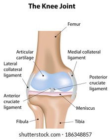
Knee joint with labels
Knee joint: anatomy, ligaments and movements | Kenhub The tibiofemoral joint Medial condyle of femur Condylus medialis femoris 1/7 The tibiofemoral joint is an articulation between the lateral and medial condyles of the distal end of the femur and the tibial plateaus, both of which are covered by a thick layer of hyaline cartilage . Anatomy of human knee joint with labels — Stock photos "Anatomy of human knee joint with labels" is an authentic stock image by StocktrekImages. It's available in the following resolutions: 1049 x 1600px, 1704 x 2600px, 3422 x 5220px. The minimum price for an image is 49$. Image in the highest quality is 3422 x 5220px, 300 dpi, and costs 449$. Similar Images Same Series Keywords Text Bones Knee Anatomy: Bones, Muscles, Tendons, and Ligaments Bones Around the Knee There are three important bones that come together at the knee joint: The tibia (shin bone) The femur (thigh bone) The patella (kneecap) A fourth bone, the fibula, is located just next to the tibia and knee joint, and can play an important role in some knee conditions.
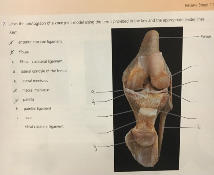
Knee joint with labels. Knee Joint - label pictures Flashcards | Quizlet Knee Joint - label pictures STUDY Flashcards Learn Write Spell Test PLAY Match Gravity Created by cfreynolds2018 Terms in this set (7) 1. Femur 2. Articular capsule 3. PCL 4. Lateral Meniscus 5. ACL 6. Tibia 1-6 7. Quadracep tendon 8. Suprapatellar bursa 9. Patella 10. Subcutaneous prepatellar bursa 11. Synovial cavity 12. Lateral Meniscus 13. Knee Joint Labeled Diagram stock vector. Illustration of arthritis ... Knee Joint Labeled Diagram stock vector. Illustration of arthritis - 39627491 Stand with Ukraine! 5% of our sales go to NGOs supporting Ukrainian causes and war refugees. More about Dreamstime Giving Fund. Our Ukrainian photographers and illustrators. Get 15 images free trial Knee Joint Labeled Diagram Royalty-Free Stock Photo Knee Joint - San Diego Mesa College Knee Joint. Click on a photo for a larger view of the model. Click on L abel for the labeled model. Back to Muscular System. Anterior: Anterior without patella: Posterior: Label: Label: Label : Label: Label: Knee Images and Pictures - Photos and X-Rays of the Knee The knee is one of the most commonly injured joints in the body. The knee joint is the junction of the thigh and the leg (part of the lower extremity). The femur (thigh bone) contacts the tibia (shin bone) at the knee joint. The patella (kneecap) sits over the front of the knee joint. Four major ligaments connect the bones and stabilize the ...
Solved Correctly label the following anatomical features of - Chegg Question: Correctly label the following anatomical features of the knee joint. Patellar ligament Synovial membrane Articular capsule Articular cartilage Fat pad Joint cavity This problem has been solved! See the answer Show transcribed image text Expert Answer 100% (2 ratings) Articular capsule. Articular … View the full answer Labeling the Knee Joint Quiz - PurposeGames.com This is an online quiz called Labeling the Knee Joint There is a printable worksheet available for download here so you can take the quiz with pen and paper. Your Skills & Rank Total Points 0 Get started! Today's Rank -- 0 Today 's Points One of us! Game Points 11 You need to get 100% to score the 11 points available Actions Add to favorites Knee Joint Anatomy: Structure, Function & Injuries - Knee Pain Exp The specific design of knee joint anatomy allows a number of functions: Supports the body in upright position without muscles having to work. Helps in lowering and raising body e.g. sitting, climbing and squatting. Allows rotation/twisting of the leg to place and position foot accurately. The Knee Joint - Articulations - Movements - TeachMeAnatomy The knee joint is a hinge type synovial joint, which mainly allows for flexion and extension (and a small degree of medial and lateral rotation). It is formed by articulations between the patella, femur and tibia. In this article, we shall examine the anatomy of the knee joint - its articulating surfaces, ligaments and neurovascular supply.
Knee x-ray - labeling questions | Radiology Case | Radiopaedia.org Normal X-ray Knee - Frontal (with labels) Annotated image Annotated image Frontal Knee Frontal 1. Femoral shaft 2. Patella 3. Base of patella 4. Apex of patella 5. Adductor tubercle of femur 6. Medial epicondyle of femur 7. Medial condyle of femur 8. Lateral epicondyle of femur 9. Lateral condyle of femur 10. Groove for popliteus 11. label the knee Quiz - PurposeGames.com This is an online quiz called label the knee. There is a printable worksheet available for download here so you can take the quiz with pen and paper. Your Skills & Rank. Total Points. 0. Get started! Today's Rank--0. Today 's Points. One of us! Game Points. 13. You need to get 100% to score the 13 points available. Knee Anatomy, Diagram & Pictures | Body Maps - Healthline Knee. The knee is a complex joint that flexes, extends, and twists slightly from side to side. The knee is the meeting point of the femur (thigh bone) in the upper leg and the tibia (shinbone) in ... Knee Joint - Anatomy Pictures and Information - Innerbody The knee, also known as the tibiofemoral joint, is a synovial hinge joint formed between three bones: the femur, tibia, and patella. Two rounded, convex processes (known as condyles) on the distal end of the femur meet two rounded, concave condyles at the proximal end of the tibia. Continue Scrolling To Read More Below... Additional Resources
34 Label Knee Joint - Labels Information List
Knee Anatomical Models | Knee Joint Models - Universal Medical Inc Ultraflx Ligamented Knee - Functional Replica. $204.00. Knee Joint with Ligaments Model. $108.00. Knee Joint with Removable Muscles 12-Part. MSRP $510.00 $469.00. Sectional Knee Joint Model 3-Part. MSRP $169.00 $156.00. Mini Knee Joint with Cross Section of Bone - On Base.

Label the parts of the knee joint. | Study.com
Knee joint Labels Flashcards | Quizlet Knee joint Labels. How do you want to study today? Flashcards. Review terms and definitions. Learn. Focus your studying with a path. Test. Take a practice test. Match. Get faster at matching terms. Created by. shelby_lemaire PLUS. Terms in this set (14) Posterior and anterior cruciate ligaments.
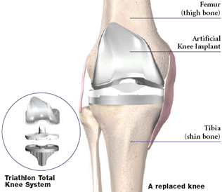
Minimally Invasive Knee Joint Replacement | Advanced Musculoskeletal ...
Knee Joint Anatomy (Labeling) Diagram - Quizlet Only $2.99/month Knee Joint Anatomy (Labeling) STUDY Learn Flashcards Write Spell Test PLAY Match Gravity Created by diegoparas Terms in this set (20) A Femur B Patellar Space Surface C Lateral Condyle D Lateral Collateral Ligament (LCL) E Lateral Meniscus F Transverse Ligament G Fibular Head H Tibial Tuberosity I Medial Condyle J

Alila Medical Media | Orthopaedics & Sport Medicine Images & Videos
knee joint label Quiz This is an online quiz called knee joint label There is a printable worksheet available for download here so you can take the quiz with pen and paper. Your Skills & Rank Total Points Get started! Today's Today 's Points Game A shoutout is a way to let people know of a game. Pick an audience - or yourself - and it'll end up in their play queue.

35 Label Knee Joint - Labels For Your Ideas
Amazon.com: anatomical model knee Axis Scientific Functional Knee Model - Anatomically Correct Knee Joint with Life Like Ligaments That Can Show Movement, Includes Base, Detailed Full Color Product Manual, Worry Free 3 Year Warranty 22 $49 99 Get it as soon as Wed, Apr 13 FREE Shipping by Amazon

Knee x-ray - Stock Image - C022/3058 - Science Photo Library
A Diagrammatic Explanation of the Parts of the Human Knee Knee actually consists of three bones - femur, tibia and patella. Femur is the thigh bone, tibia is the shin bone and patella is the small cap like structure which rests on the other two bones. Femur is considered as the largest bone in the human body. The femur and the tibia meets at the tibiofemoral joint and patella rests on top of this joint.

X-rays Lower limb
Knee Joint Model Labeling Quiz - PurposeGames.com This is an online quiz called Knee Joint Model Labeling Quiz There is a printable worksheet available for download here so you can take the quiz with pen and paper. Your Skills & Rank Total Points 0 Get started! Today's Rank -- 0 Today 's Points One of us! Game Points 15 You need to get 100% to score the 15 points available Actions

Easing joint pain - The science behind supplements - DannyUK - The ...
A Labeled Diagram of the Knee With an Insight into Its Working Labeled Diagram of the Knee Joint Knee joint is one of the most important hinge joints of our body. Its complexity and its efficiency is the best example of God's creation. The anatomy of the knee consists of bones, muscles, nerves, cartilages, tendons and ligaments. All these parts combine and work together.

Knee Joint Replacement, Illustration - Stock Image - F031/7379 ...
Knee Joint Label Flashcards | Quizlet Knee Joint Label STUDY Flashcards Learn Write Spell Test PLAY Match Gravity Created by LaLaKub91 Terms in this set (10) femur What is A? lateral collateral ligament what is d? lateral meniscus what is e? fibula what is g? tibia what is h? posterior cruciate ligament What is j? anterior cruciate ligament what is k? medial meniscus what is l?
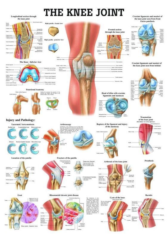
The Knee Joint Laminated Anatomy Chart | Knee joint anatomy, Joints ...
The knee (MRI): Atlas of anatomy in medical imagery - IMAIOS Anatomy of the knee on a coronal slice (MRI) : meniscus (lateral and medial), cruciate ligaments, vastus (lateralis, intermedius, medialis), tibial and fibular collateral ligaments. On "Contrast" the user can choose the type of MRI sequence: spin-echo T1 or proton-density with fat saturation sequences. On "Series" it is possible to ...

Radiology Cases: Synovial Hypertrophy
Knee Anatomy: Bones, Muscles, Tendons, and Ligaments Bones Around the Knee There are three important bones that come together at the knee joint: The tibia (shin bone) The femur (thigh bone) The patella (kneecap) A fourth bone, the fibula, is located just next to the tibia and knee joint, and can play an important role in some knee conditions.

Bicep Tendon Sprain Front Shoulder Pain Sioux City
Anatomy of human knee joint with labels — Stock photos "Anatomy of human knee joint with labels" is an authentic stock image by StocktrekImages. It's available in the following resolutions: 1049 x 1600px, 1704 x 2600px, 3422 x 5220px. The minimum price for an image is 49$. Image in the highest quality is 3422 x 5220px, 300 dpi, and costs 449$. Similar Images Same Series Keywords Text Bones

Exercise Anatomy for Students: April 2009
Knee joint: anatomy, ligaments and movements | Kenhub The tibiofemoral joint Medial condyle of femur Condylus medialis femoris 1/7 The tibiofemoral joint is an articulation between the lateral and medial condyles of the distal end of the femur and the tibial plateaus, both of which are covered by a thick layer of hyaline cartilage .

Alila Medical Media | Knee joint diagram unlabeled | Medical illustration
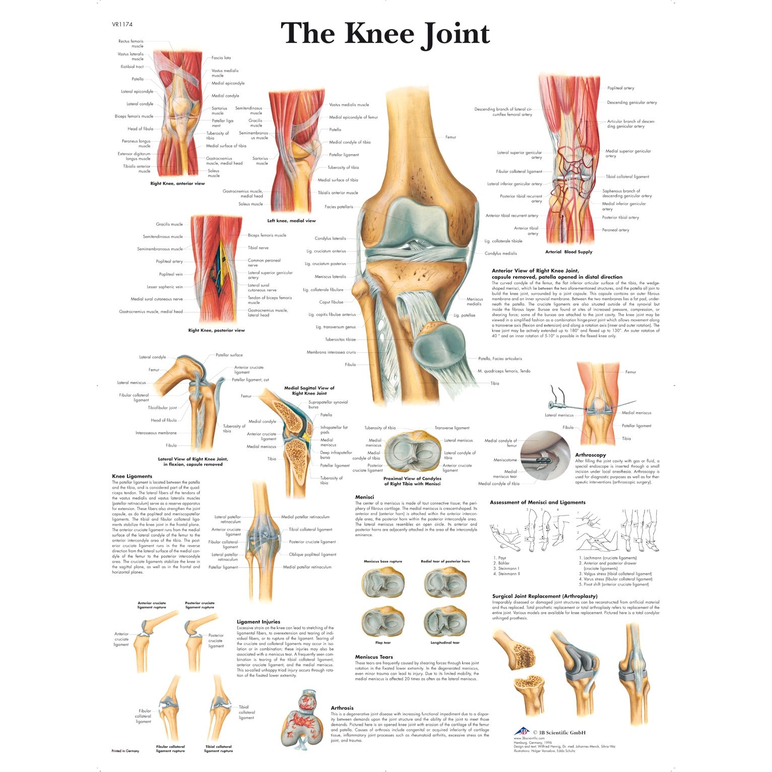
3B Scientific The Knee Joint Chart - English

Knee joint features - YouTube
30 Label The Knee - Labels Information List

Synovial Joint
Post a Comment for "40 knee joint with labels"