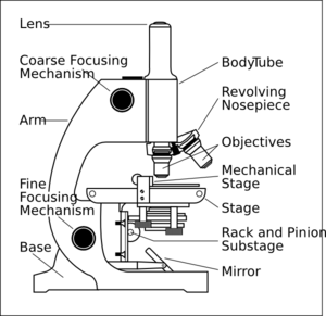
45 compound microscope diagram without labels
EOF Electron Microscope: Principle, Types, Applications - Microbe Online Electron microscopes are used for detailed investigation of the ultrastructure of a wide range of biological and inorganic specimens including microorganisms, cells, large molecules, biopsy samples, metals, and crystals. German physicist Ernst Ruska invented electron microscope in 1931. Components of an Electron Microscope
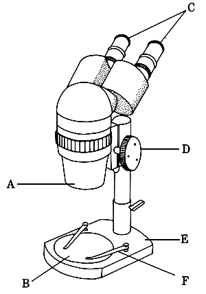
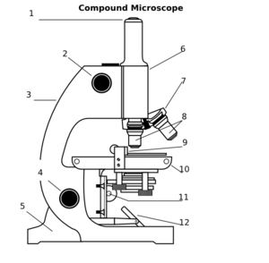
Light Microscope (Theory) - Amrita Vishwa Vidyapeetham The modern compound microscope consists of two lens system, the objective and the ocular or eye piece. The first magnified image obtained with objective lens, is again magnified by the eye piece to give a virtual inverted image. The total magnification the product of the magnifications of two lens systems. Parts of a Microscope

Compound microscope diagram without labels
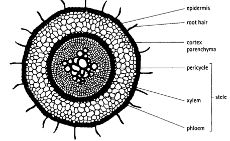
Mitosis in Onion Root Tips - Amrita Vishwa Vidyapeetham Then the thin layer of cell squash on the slide was viewed under the light microscope. Then the cell was photographed and documented. By using actively dividing cells in the onion root tip, this experiment aims to obtain a karyotype from the sample and to determine the purpose of each step used in the procedure.
Compound microscope diagram without labels. Mitosis in Onion Root Tips - Amrita Vishwa Vidyapeetham Then the thin layer of cell squash on the slide was viewed under the light microscope. Then the cell was photographed and documented. By using actively dividing cells in the onion root tip, this experiment aims to obtain a karyotype from the sample and to determine the purpose of each step used in the procedure.
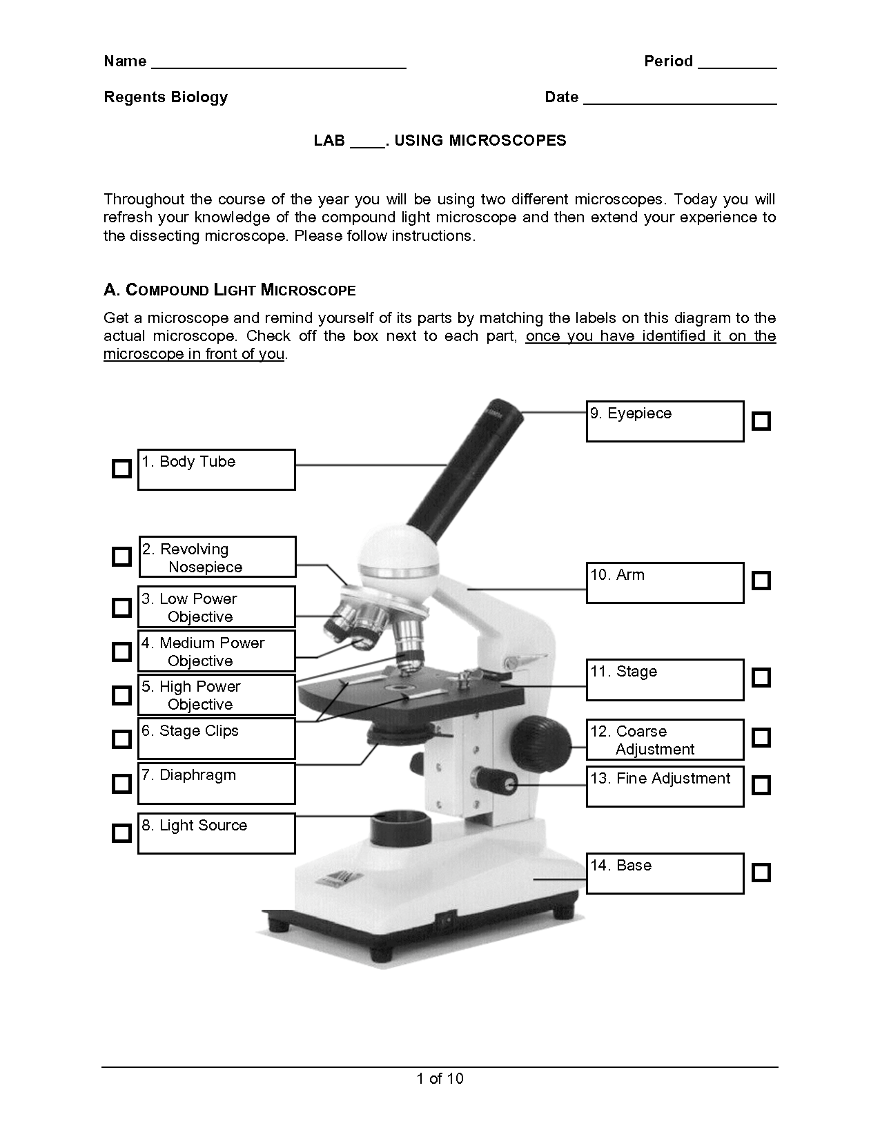
8 Best Images of Using A Microscope Worksheet - Compound Microscope ...
Microscope With Labels Clip Art at Clker.com - vector clip art online ...

Microscope With Labels Clip Art at Clker.com - vector clip art online ...
Sketch Microscope Diagram Easy To Draw - Micropedia
16 Best Images of Simple Microscope Labeling Worksheet - Compound Light ...

Microscope With Labels Clip Art at Clker.com - vector clip art online ...
Compound Microscope Clip Art at Clker.com - vector clip art online ...
16 Best Images of Simple Microscope Labeling Worksheet - Compound Light ...

Compound Microscope Drawing - Micropedia
Post a Comment for "45 compound microscope diagram without labels"