41 onion cells under microscope with labels
Observing Onion Cells Under The Microscope Afterwards, carefully mount the prepared and stained onion cell slide onto the microscope stage. Make sure that the cover slip is perfectly aligned with the microscope slide, and that any excess stain has been wiped off. Secure the slide on the stage using the stage clips. Onion Epidermal Cell Labeled Diagram - schematron.org Nov 15, 2018 · Draw a labelled diagram of an onion epidermal cell seen under the microscope. ( 4 marks) e The onion epidermal cells are not green in colour because they lack. The epidermal cells of onions provide a protective layer against viruses and fungi that may harm the sensitive tissues.
BIOLOGY EXPERIMENT EXAMINATION OF ONION CELL IN LIGHT MICROSCOPE Aug 20, 2019 · Place the single layer of onion cell epithelium on a glass slide. Make sure that you do not fold it over or wrinkle it. Place a drop of iodine stain on your onion tissue. Put the cover slip on the stained tissue and gently tap out any air bubbles. Observe the cells under 4x, 10x, and 40x with the diaphragm wide open.
Onion cells under microscope with labels
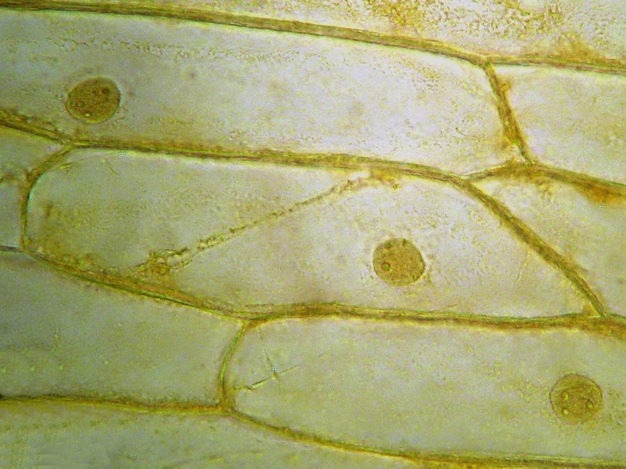
Onion Cells Under a Microscope - Requirements/Preparation ... An onion is made up oflayers that are separated by a thin membrane. For this experiment, the thinmembrane will be used to observe the onion cells. It can easily be obtained bypeeling it from any layer of the onion using tweezers. Onion Cells - Investigation - Exploring Nature 5. Observe the onion tissue under the microscope at 4x, 10x and 40x with lots of light (open diaphragm). Then slowly close the diaphragm while observing the image to find the best light for seeing cellular details. 6. Draw a section of onion skin cells at 10x magnification. Then switch to 40x and draw one cell and label it. Questions: 1.
Onion cells under microscope with labels. Onion Cells - Investigation - Exploring Nature 5. Observe the onion tissue under the microscope at 4x, 10x and 40x with lots of light (open diaphragm). Then slowly close the diaphragm while observing the image to find the best light for seeing cellular details. 6. Draw a section of onion skin cells at 10x magnification. Then switch to 40x and draw one cell and label it. Questions: 1. Onion Cells Under a Microscope - Requirements/Preparation ... An onion is made up oflayers that are separated by a thin membrane. For this experiment, the thinmembrane will be used to observe the onion cells. It can easily be obtained bypeeling it from any layer of the onion using tweezers.
The inner epidermis of the onion bulb’s cataphylls (the onion skin).
Post a Comment for "41 onion cells under microscope with labels"